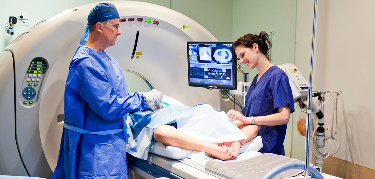
Ultrasound uses high-frequency sound waves to create detailed images of the inside of the body. Sometimes referred to as sonography, the machine involves an advanced computer connected to a handheld transducer, which is a combined sound transmitter and microphone that sends sound waves into your body above the human range of hearing. The device then picks up the echoes from your tissue; different types of tissue produce echoes of different types. In addition, injured or diseased tissue can produce a different echo from healthy tissue. The computer receives and processes this information in order to create a detailed image for interpretation by a radiologist.
Ultrasound is often used to examine the abdomen to evaluate infections, tumors or inflammation. It is commonly used to examine women’s pelvic areas, evaluate pregnancies and detect gallstones; it is useful in looking for cysts or tumors.


